| Sur un cas d'agrypnie (4 mois sans sommeil) au cours d'une maladie de Morvan. Effet favorable du 5-hydroxytryptophane |
| C. Fischer-Perroudon, J. Mouret et M. Jouvet Electroencephalography and Clinical Neurophysiology, 1974, 36: 1-18 |
| IMPRESSION |
| Version imprimable (Tout l'article dans une seule page) |
Figure 7 : Records taken during hallucinatory bebaviour associated with distal pain
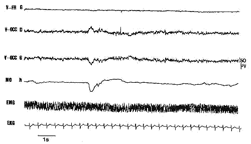
Derivations between vertex and left frontal region, right
and left occipItal. regions.
Electro-oculogram (horizontal), suprahyoid EMG activity and EKG.
Calibration: 1 sec, 50 microV