| Iontophoretic application of unconjugated cholera toxin B subunit (CTb) combined with immunohistochemistry of neurochemical substances: a method for transmitter identification of retrogradely labeled neurons |
| Luppi P.H., Fort P., Jouvet M. Brain Res. 534 (1-2) pages : 209-224 (1990) |
| TABLE OF CONTENTS |
| Materials and Methods |
| Results |
| Figures |
| Printable version |
Figure 5
Click on photomicrographs A,B,C or D to get it bigger
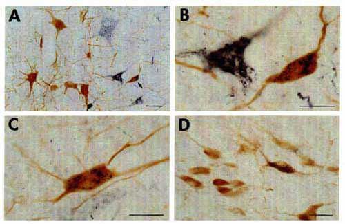
A-D: color photomicrographs illustrating CTb-labeled cells charaeterized hy black punctate labeling and AADC-(A-C) and ChAT (D)-immunoreactive cells characterized by diffuse light brown staining. Double-labeled cells are easily distinguished in the nucleus raphe magnus (A-C) and the pedunculopontine nucleus (D) in the pons. The arrow in A indicates the double-labeled cell shown at a higher magnification in B.
Bars = 50 µm (A) and 25 µm (B-D).
Nomenclature according to Berman 4.