| Iontophoretic application of unconjugated cholera toxin B subunit (CTb) combined with immunohistochemistry of neurochemical substances: a method for transmitter identification of retrogradely labeled neurons |
| Luppi P.H., Fort P., Jouvet M. Brain Res. 534 (1-2) pages : 209-224 (1990) |
| TABLE OF CONTENTS |
| Materials and Methods |
| Results |
| Figures |
| Printable version |
Figure 3E
A: overview of a section at pontine level
showing a large iontophoretic injection site (2 uA, 60 min) restricted
to the nucleus raphe dorsalis in a cat with 18 days of survival (case
R121) The other photomicrographs of this figure illustrate examples of
retrograde and anterograde labeling obtained in this case.
Bar = 2 mm.
B: low power photomicrograph showing
the presence of numerous retrogradely labeled cells in the lateral habenular
nucleus, indicating that the quantity of CTb in these retrogradely labeled
cells rather than decreasing, increased after 18 days of survival in this
well-known afferent to the nucleus raphe dorsalis.
Bar = 100 µm.
C,D: photomicrographs
of retrogradely labeled neurons in the basal forebrain (C) and the insular agranular cortex (D). Note the very extensive labeling of the primary and
secondary dendritic branches in the neuron shown in C and of the large dendrites of the pyramidal cells shown
in D.
Bar = 50 µm.
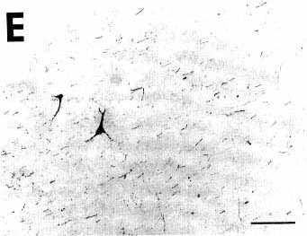
E: photomicrograph illustrating in the
median forebrain bundle the presence of two retrogradely labeled cell
bodies surrounded by the bundle of anterogradely labeled fibers arising
from the nucleus raphe dorsalis. Note that these fibers are passing and
therefore do not display varicosities.
Bar = 100 µm.
F: photomicrograph illustrating the presence
of numerous anterogradely labeled fibers with terminal-like swellings
in the superficial layers of the caudal part of the pyriform cortex.
Bar = 50 µm.