| Coma and other disorders of consciousness |
| Jouvet M. Handbook of Clinical Neurology Vol.3. P. J. Vinken and G. W. Bruyn , eds. North-Holland Publishing Company. Amsterdam,(1969) |
| TABLE OF CONTENTS |
|
Periodic physiological dissolution of consciousness: sleep and coma |
|
Aetiological classification of comas and of disturbances of consciousness of organic origin |
| FIGURES |
| Printable version |
Figure 8 : Diagrammatic anatomoclinical correlation of main forms of coma
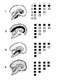
On the left: the most common lesions are shown in black on a
sagittal section of the encephalon; less frequent lesions shown as dotted
areas.
On the right: results of the clinical examination. Black squares
indicate a negative response to each test.
- (1) Akinetic mutism: P3-D3-R1-VI.
- (2) Decortication syndrome: P5-D1-RI-VI.
- (3) Upper brainstem lesion: P5-D3-R2 Vl.
- (4) Brain death: P5-D4-R3-V2. See details in text.