|
Afferent projections to the rat locus coeruleus demonstrated by retrograde and anterograde tracing with cholera-toxin B subunit and Phaseolus vulgaris leucoagglutinin |
| Luppi P.H., Aston-Jones G., Akaoka H., Chouvet and Jouvet M. Neuroscience (1995) Vol:65 Tome:1 pp:119-160 |
| TABLE OF CONTENTS |
| RESULTS |
|
II. Afferent projections to the locus coeruleus |
| FIGURES |
Figure 6B
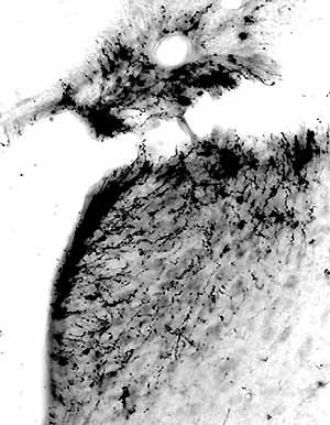
Photomicrograph showing anterogradely labeled fibers in the nuclear core of the rat LC after the PHAL injection in the lateral part of the periaqueductal gray shown in D. Only occasional fibers are localized in the MVe region lateral to the LC. Note that the dorsal LC is partly obscured by artefactual labeling in a tear in the tissue.