|
Afferent projections to the rat locus coeruleus demonstrated by retrograde and anterograde tracing with cholera-toxin B subunit and Phaseolus vulgaris leucoagglutinin |
| Luppi P.H., Aston-Jones G., Akaoka H., Chouvet and Jouvet M. Neuroscience (1995) Vol:65 Tome:1 pp:119-160 |
| TABLE OF CONTENTS |
| RESULTS |
|
II. Afferent projections to the locus coeruleus |
| FIGURES |
Figure 24AB
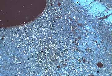
Figure 24a
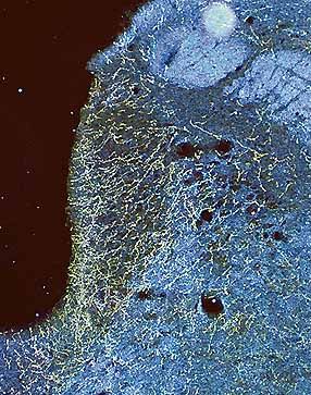
Figure 24b
Color darkfield photomicrographs of frontal sections respectively at the level of Barrington's nucleus (A) and the LC (B) showing the presence of anterogradely labeled fibers in these nuclei after the PHAL injection in the nucleus Kolliker-Fuse shown in Fig. 5B