|
Afferent projections to the rat locus coeruleus demonstrated by retrograde and anterograde tracing with cholera-toxin B subunit and Phaseolus vulgaris leucoagglutinin |
| Luppi P.H., Aston-Jones G., Akaoka H., Chouvet and Jouvet M. Neuroscience (1995) Vol:65 Tome:1 pp:119-160 |
| TABLE OF CONTENTS |
| RESULTS |
|
II. Afferent projections to the locus coeruleus |
| FIGURES |
Figure 23E
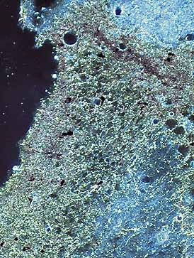
Darkfield color photomicrograph illustrating anterogradely labeled fibers in the LC and surrounding structures after a CTb injection in the perifornical nucleus dorsal to the fornix at the level of the dorsomedial hypothalamic nucleus. Note the terminal-like dots in the nuclear core of the LC. More terminal-like labeling is visible in the medial parabrachial nucleus and the lamina between the LC and the fourth ventricle.