|
Afferent projections to the rat locus coeruleus demonstrated by retrograde and anterograde tracing with cholera-toxin B subunit and Phaseolus vulgaris leucoagglutinin |
| Luppi P.H., Aston-Jones G., Akaoka H., Chouvet and Jouvet M. Neuroscience (1995) Vol:65 Tome:1 pp:119-160 |
| TABLE OF CONTENTS |
| RESULTS |
|
II. Afferent projections to the locus coeruleus |
| FIGURES |
Figure 23CD
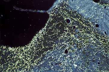
Figure 23c
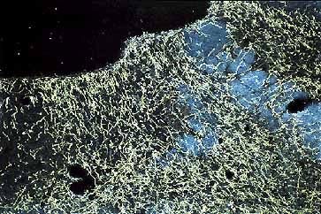
Figure 23d
Darkfield color photomicrograph of frontal sections illustrating anterogradely labeled fibers in the LC in C and in the nuclei rostral to it in D after a PHAL injection in the lateral hypothalamic area of the posterior hypothalamus. The injection site was localized dorsolaterally to the fornix at the rostral level of the subthalamic nucleus. Note in C the fibers localized in the nuclear core of the LC. More fibers are found in surrounding structures. In D, the anterogradely labeled fibers are diffusely distributed over the Bar, peri5Me, 5Me, rostral pole of the LC, medial parabrachial nucleus and the periaqueductal gray surrounding these nuclei.