|
Afferent projections to the rat locus coeruleus demonstrated by retrograde and anterograde tracing with cholera-toxin B subunit and Phaseolus vulgaris leucoagglutinin |
| Luppi P.H., Aston-Jones G., Akaoka H., Chouvet and Jouvet M. Neuroscience (1995) Vol:65 Tome:1 pp:119-160 |
| TABLE OF CONTENTS |
| RESULTS |
|
II. Afferent projections to the locus coeruleus |
| FIGURES |
Figure 22D
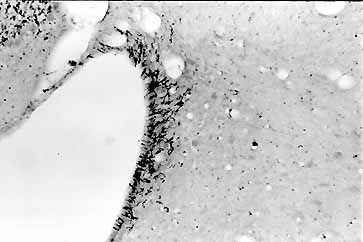
Photomicrograph illustrating anterogradely labeled fibers in the ependyma and the extreme caudal tip of the LC after the PHAL injection in the preoptic region shown in E. Note that the MVe area lateral to the LC contained no labeled fibers.