| Iontophoretic application of unconjugated cholera toxin B subunit (CTb) combined with immunohistochemistry of neurochemical substances: a method for transmitter identification of retrogradely labeled neurons |
| Luppi P.H., Fort P., Jouvet M. Brain Res. 534 (1-2) pages : 209-224 (1990) |
| TABLE OF CONTENTS |
| Materials and Methods |
| Results |
| Figures |
| Printable version |
Figure 2
Click on photomicrographs A,B,C,D,E or F to get it bigger
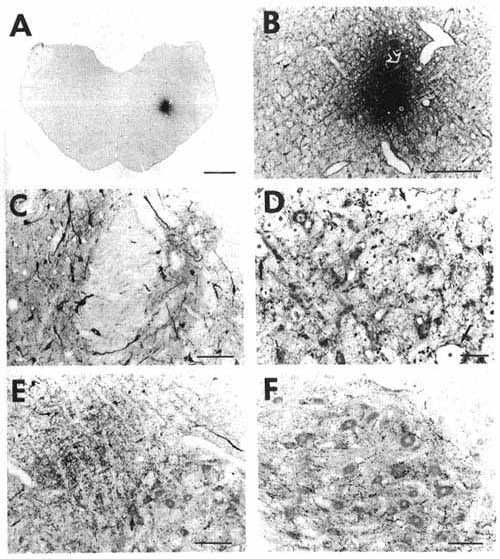
A: overview of a frontal section at the
level of the medulla oblongata showing a typical iontophoretic injection
site in the nucleus reticularis parvicellularis (case K120) using a 2-µA
pulsed positive current for 30 min.
Bar= 2 mm.
B: higher magnification of the site shown
in A precisely illustrating its extent and localization. The stars and
arrows designate the same vessels or fiber bundle in B and D.
Bar = 400 µm.
D: adjacent section to that shown in
B at a higher magnification, counterstained with Cresyl violet and immunostained
with serotonin, showing the presence of counterstained cell bodies and
serotonin immunoreactive fibers in the site proving the absence of tissue
necrosis.
Bar =50 µm.
C: photomicrograph illustrating retrogradely
labeled cells in the medial (left part) and lateral (right part) divisions
of the nucleus of the solitary tract. Note also the presence of punctate
anterograde labeling in the same areas.
Bar = 100 µm.
E: photomicrograph showing anterogradely
labeled fibers organized as a bundle running dorsally to the facial nucleus
approximately 2 mm rostral to the site shown in A
and B. Varicose fibers inside the facial nucleus
sometimes formed basket-like structures around motoneuron somata (arrow).
Bar = 200 µm.
F: photomicrograph showing the great
number of anterogradely labeled varicose fibers in the hypoglossal nucleus
after the CTb iontophoretic injection shown in A
and B.
Bar = 100 µm.
To optimize the CTb immunostaining shown in C, E and F, the sections were incubated 3-4 days in CTb antiserum (1: 40,000) at 4 °C followed by sequential incubations overnight at 4 °C in the biotinylated donkey anti-goat IgG (1:2,000) and the streptavidin-HRP (1:40,000). Note that with such maximal conditions, we obtained a weak counterstaining of the sections ideally suited for accurate localization of CTb immunostained material.