| A Study of the Neurophysiological Mechanisms of Dreaming |
| M. Jouvet and D. Jouvet Electroenceph. Clin. Neurophysiol. 1963 Suppl. 24 |
| TABLE OF CONTENTS |
| Part 1 |
| Part 2 |
| Figures |
| Printable version |
Fig. 8 : Diagrammatic representation of the sleep-wakefulness rhythm
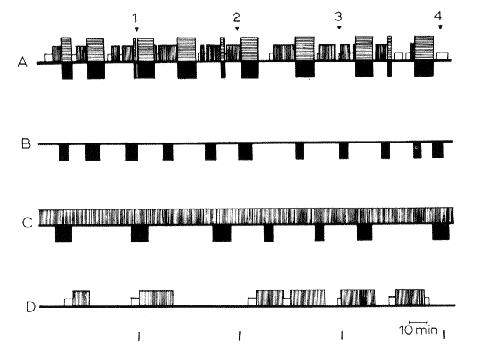
in (A) intact, (B) decorticate, (C) mesencephalic cats, and (D) cats with coagulation of the nucleus reticularis pontis caudalis. 4 h of continuous recordings were taken in each cat. In black: RPS with fast cortical EEG pattern in normal cats (horizontal hatching) and spindling activity at the pontine level, total disappearance of EMG activity, eye movements, in normal, decorticate and mesencephalic cats. In white: spindling activity on the cortex. Verfical hatching: slow waves and spindles at the cortical and diencephalic level. Horizontal line: arousal. Note the absence of SPS in decorticate cats, and the absence of RPS in the cat with pontine lesion. The slow EEG activity in C represents the "lethargic state of the brain "in cerveau isole' but not a true "slow sleep".
Time scale: 10 min.
From Jouvet (1961), with kind permission of J. and A. Churchill Ltd., London.