| A Study of the Neurophysiological Mechanisms of Dreaming |
| M. Jouvet and D. Jouvet Electroenceph. Clin. Neurophysiol. 1963 Suppl. 24 |
| TABLE OF CONTENTS |
| Part 1 |
| Part 2 |
| Figures |
| Printable version |
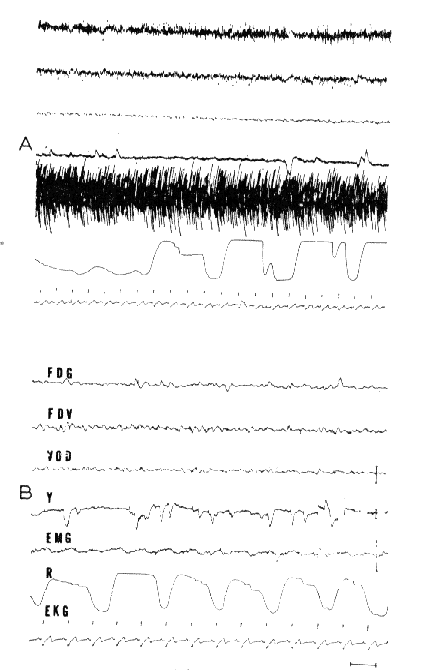
Fig. 17 : Arousal and RPS in a syndrome of decortication

A. Arousal: Very low voltage EEG activity and considerable EMG activity at the frontal region and the biceps (hypertony of decortication). Periodic breathing.
B. RPS: Total disappearance of the EMG activity. Discrete rhythmic 3/sec slow waves at the vertex. Rapid eye movements (Y). Slowing of the EKG. Acceleration of respiration.
- FDG: right and left frontal;
- FDV: vertex-right frontal;
- VOD: vertex-right occipItal.
Scale: 1 sec, 50 microV.