| Telencephalic and rhombencephalic sleep in the cat |
| Jouvet M. The Nature of Sleep Ciba Foundation Symposium Churchill (1961) |
| TABLE OF CONTENTS |
|
Topography of the systems responsible for the two stages of sleep |
| Figures |
| Printable version |
Figure 7
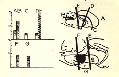
Diagrammatic representation of the sleep-wakefulness rhythm in (A) intact, (B) cerebellectomized, (D) mesencephalic, (E) pontine, (F) retropontine cats, and (G) in cats with limited medial lesion of the pontine reticular formation (in black) (cf. Fig. 8).
Numbers at left indicate the percentage, for 12 hours continuous recording, of either synchronized and slow activity (columns with dots) or fast activity and disappearance of EMG activity during sleep (white columns). Bach category represents the mean of sixteen recordings in four cats. Cross-hatching indicates standard deviation. Note the absence of synchronized activity in decorticate cat (C), and the absence of the rhombencephalic stage of sleep in F and G. In mesencephalic and pontine cats (D, E), the rhombencephalic stage of sleep does not induce fast activity at the cortical level and is distinguished on the BMG and by the pontine activity.
N.R.: Red nucleus.
P: Pons.
C. Tr.: Trapezoid body.