|
Afferent projections to the rat locus coeruleus demonstrated by retrograde and anterograde tracing with cholera-toxin B subunit and Phaseolus vulgaris leucoagglutinin |
| Luppi P.H., Aston-Jones G., Akaoka H., Chouvet and Jouvet M. Neuroscience (1995) Vol:65 Tome:1 pp:119-160 |
| TABLE OF CONTENTS |
| RESULTS |
|
II. Afferent projections to the locus coeruleus |
| FIGURES |
Figure 5E
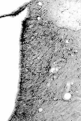
Photomicrograph showing anterogradely labeled fibers in the LC after the PHAL injection in the nucleus Kšlliker-Fuse shown in B. Note also the presence of a large number of fibers in the MVe area just lateral to the LC and in the lamina between the LC and the fourth ventricle.