|
Afferent projections to the rat locus coeruleus demonstrated by retrograde and anterograde tracing with cholera-toxin B subunit and Phaseolus vulgaris leucoagglutinin |
| Luppi P.H., Aston-Jones G., Akaoka H., Chouvet and Jouvet M. Neuroscience (1995) Vol:65 Tome:1 pp:119-160 |
| TABLE OF CONTENTS |
| RESULTS |
|
II. Afferent projections to the locus coeruleus |
| FIGURES |
Figure 24CD
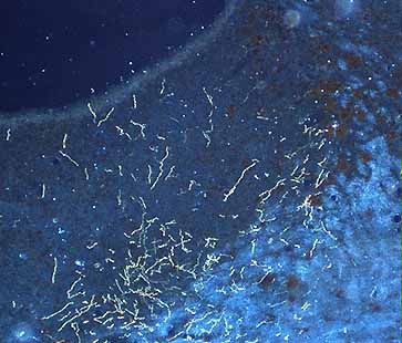
Figure 24c
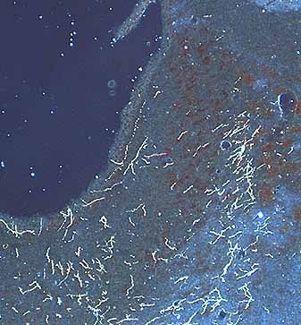
Figure 24d
Color darkfield photomicrographs of frontal sections showing the distribution of anterogradely labeled fibers at the level of Barrington's nucleus (C) and the LC (D) after a PHAL injection in the area 1 of the frontal cortex. Note that only a few anterogradely labeled fibers are localized in the LC itself.