|
Afferent projections to the rat locus coeruleus demonstrated by retrograde and anterograde tracing with cholera-toxin B subunit and Phaseolus vulgaris leucoagglutinin |
| Luppi P.H., Aston-Jones G., Akaoka H., Chouvet and Jouvet M. Neuroscience (1995) Vol:65 Tome:1 pp:119-160 |
| TABLE OF CONTENTS |
| RESULTS |
|
II. Afferent projections to the locus coeruleus |
| FIGURES |
Figure 23AB
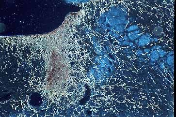
Figure 23a
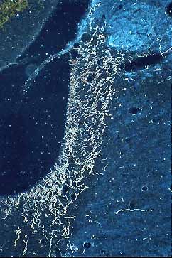
Figure 23b
Darkfield color photomicrograph showing anterogradely labeled fibers in the Bar (A) and the LC (B) after the PHAL injection in the preoptic area shown in Fig. 22E. A dense plexus of fibers is covering the Bar in A. Fibers are localized in the nuclear core of the LC in B. Note that only few fibers are localized in the MVe region just lateral to the LC.