| A Study of the Neurophysiological Mechanisms of Dreaming |
| M. Jouvet and D. Jouvet Electroenceph. Clin. Neurophysiol. 1963 Suppl. 24 |
| TABLE OF CONTENTS |
| Part 1 |
| Part 2 |
| Figures |
| Printable version |
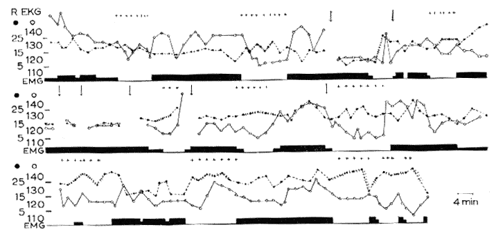
Fig. 18 : Sleep-wakefulness rhythm in a syndrome of decortication

Continuous recordings during 8 h (from 9 p.m. to 5 a.m.). Vertical arrows signal brief interruption of the recording.
Abscissa: In black: EMG activity (biceps).
Black dots and interrupted line: respiration (per min). The irregularity of respiration is marked by small vertical lines on the respiratory curve.
White dots and solid line: Heart rate (per min).
Crosses: rapid eye movements.
Ordinate: Respiratory and heart rate (per min). Note the periodical appearance of RPS (25 per cent of the time) during which there are REM, cardiorespiratory variations and a total disappearance of the EMG.