| A Study of the Neurophysiological Mechanisms of Dreaming |
| M. Jouvet and D. Jouvet Electroenceph. Clin. Neurophysiol. 1963 Suppl. 24 |
| TABLE OF CONTENTS |
| Part 1 |
| Part 2 |
| Figures |
| Printable version |
Fig. 10 : Schematic representation of the neural structures responsible for RPS
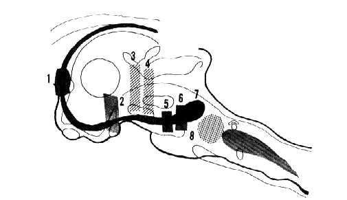
In dots (8). Nucleus reticularis pontis caudalis whose destruction suppresses RPS. In black. An ascending part of the limbic midbrain circuit with the "limbic midbrain area" of Nauta and Kuypers (1957). 1-2-5-6. Lesions of the septum, subthalamic region, interpeduncular region, and medial part of the anterior pontine tegmentum. These lesions suppress, totally or in part, the fast cortical activity and the theta hippocampal rhythm during RPS. 3-4. Lesions interrupting the ascending reticular activating system at the mesencephalic level. These lesions, which suppress cortical arousal, do not eliminate the possibility of a fast cortical activity during RPS. In grey. Ponto-bulbar inhibitory reticular formation which is probably responsible for the total atony during RPS.
After Jouvet (1962), with kind permission of Arch. Ital. Biol. (full-text)